flow cytometry results explained
November 3 2021 Press Release. The collaboration with Thermo Fisher Scientific will provide a full workflow from sample to results for scientists performing multiplexed single cell proteomics analyses.
Blog Flow Cytometry Data Analysis I What Different Plots Can Tell You
Polyclonal increase in any or all of the three classes IgG IgA andor IgM Infections acute and chronic including HIV Epstein-Barr virus cytomegalovirus Connective tissue diseases rheumatoid arthritis systemic lupus.
. CAD most often results from a condition known as atherosclerosis which happens when a waxy substance forms inside the arteries that supply blood to your heart. Total splenocytes harvested on ay 28 D and 121. Re-stimulated with the -length S peptide pool full high levels of secreted.
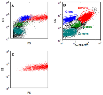
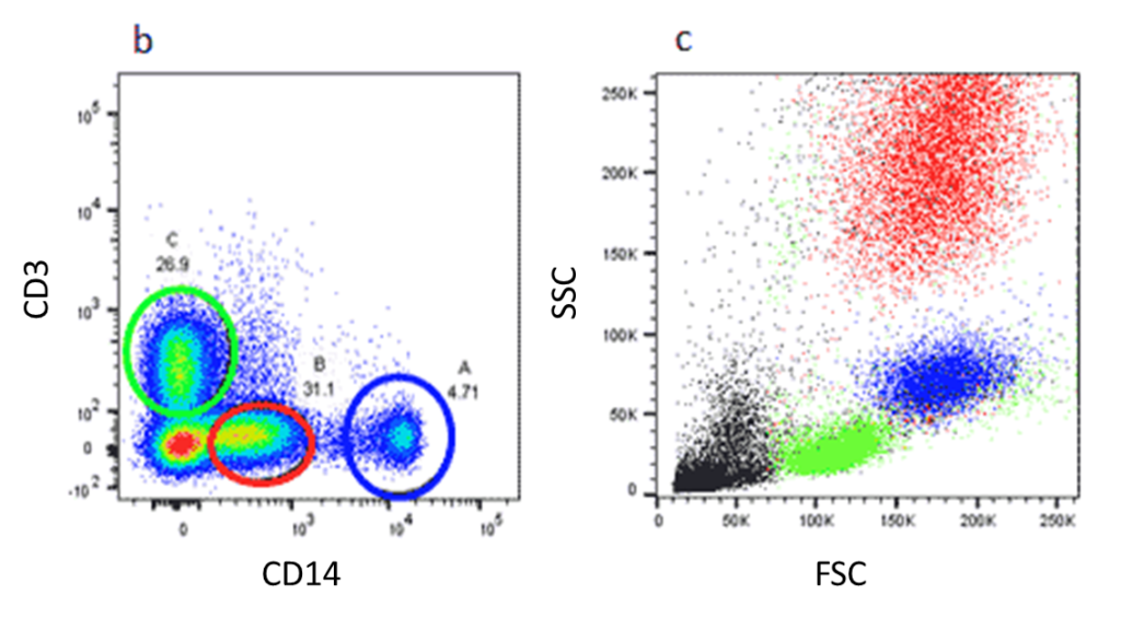
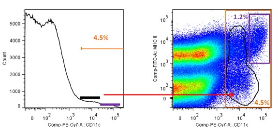
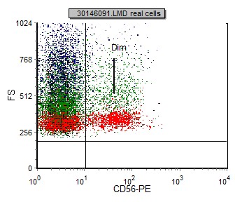
This flow cytometry guide aims to give you a basic overview of all the important aspects of flow cytometry. Causes of increased immunoglobulin levels. In this case k m for all practical situations can be described by 8 19 where D is the diffusion coefficient h and b are the height and the width of the flow cell respectively v is the volumetric flow rate and x is the distance from the flow cell entrance.
With a fulllength S peptide pool- Fig. 119 in ELISpot and intracellular cytokine staining flow cytometry analysis when restimulated - ex 120 vivo. 142 Calibration-Free Concentration Analysis CFCA.
Immunoglobulin result Associated conditions. Flow cytometry data were acquired on a BD LSR Fortessa TM cell analyzer Becton Dickinson Franklin Lakes NJ and analyzed using FlowJo software FlowJo LLC. The primary role of the Facility is to apply state of the art techniques in mass spectrometry to address important biological questions within the remit of the Institute READ.
As the plaque builds up the artery narrows making it more difficult for blood to flow to the heart. With chapters on instrumentation useful reagents controls experimental set up and much more this guide enables best practice to be followed and gives practical advice on building multicolor panels with example protocols. These causes are further explained in Table 2.
The Babraham Institute Flow Cytometry Core Facility offers high quality service and state-of-the-art instrumentation to members of Babraham Institute and external users.

Flow Cytometry Basics Flow Cytometry Miltenyi Biotec Technologies Macs Handbook Resources Miltenyi Biotec Usa
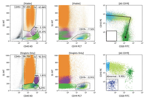
Hematologic Diagnostics Go With The Flow

Introduction To Flow Cytometric Analysis Flow Cytometry
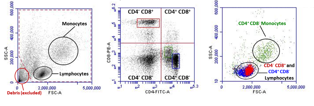
Cell Population Flow Cytometry Analysis Sygnature Discovery

Quantitative Flow Cytometry Measurements Nist
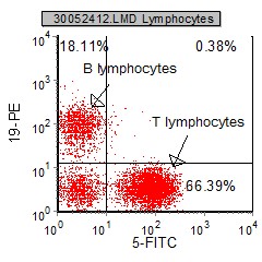
Introduction To Flow Cytometric Analysis Flow Cytometry

Show Dot Blot Analysis Of Flow Cytometry Data Of Cd4 Cd8 Of Two Cases Download Scientific Diagram

Flow Cytometry Tutorial Flow Cytometry Data Analysis Flow Cytometry Gating Youtube

What Is Flow Cytometry Facs Analysis

Dot Plot Representing Four Quadrant Images Observed By Flow Cytometric Download Scientific Diagram

Chapter 4 Data Analysis Flow Cytometry A Basic Introduction

Typical Data From A Two Color Flow Cytometry Experiment To Measure Cell Download Scientific Diagram

Overview Of High Dimensional Flow Cytometry Data Analysis A Fcs Download Scientific Diagram

Flow Cytometric Immunophenotyping Performed On The Same Plasmablastic Download Scientific Diagram

Flow Cytometry Analysis Of Cellular Uptake A The Sample Stream Download Scientific Diagram

Flow Cytometry And The Sheath Fluid You Use Lab Manager

Overview Of High Dimensional Flow Cytometry Data Analysis A Fcs Download Scientific Diagram
